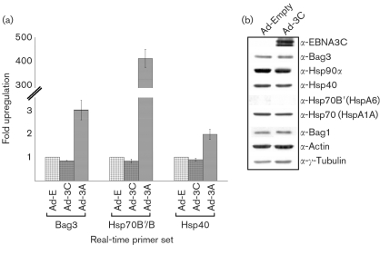
Fig. 3.
No increase in chaperones or co-chaperones was observed after infection with adenovirus EBNA3C. (a) Real-time qRT-PCR analysis with primers for Bag3, Hsp40 and Hsp70B/B′. No significant increase in Bag3, Hsp40 or Hsp70B/B′ was observed with Ad-3C (m.o.i. of 25, 24 h) compared with Ad-E control infection (Ad-3A infection results are included for comparison). GAPDH is run as an endogenous control to which all samples are normalized. (b) Western blot analysis of protein samples taken from IMR-90 cells infected with Ad-3C versus Ad-E control infection. No increase in chaperones or co-chaperones was seen with EBNA3C expression.

