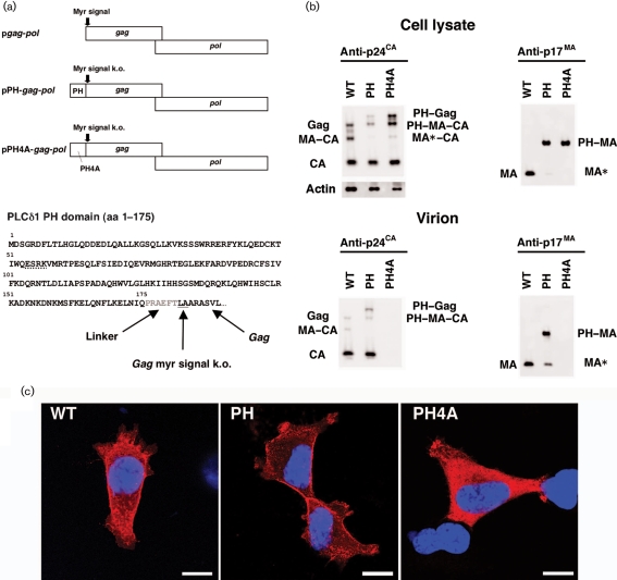
Fig. 1.
Viral production by the gag–pol expression vectors. (a) The genetic structure of the pgag-pol, pPH-gag-pol and pPH4A-gag-pol expression vectors, and the amino acid sequence of the PH–Gag junction are shown. The N-terminal PH domain of PLCδ1 (aa 1–175) was fused to LA–Gag, linked by a 5 aa spacer (shown in grey). The MG→LA mutation to knock out the myristoylation signal of Gag (myr signal k.o.) is underlined. Four alanine mutations were introduced to replace the ESRK sequence (dotted line) to create the PH4A mutant. (b) Protein expression from pgag-pol (WT), pPH-gag-pol (PH) and pPH4A-gag-pol (PH4A) in transfected 293T cell lysates and Gag cleavage in the virions were examined by Western blot analysis using anti-p24CA or anti-p17MA antibodies. Note that the anti-p17MA antibody recognizes the cleaved p17MA protein only. The band denoted as PH–MA–CA in the virion detected by the anti-p24CA antibody (lower left panel) possibly overlaps with a faint Gag signal derived from PH–Gag and PH4A–Gag from which the PH and PH4A domains have been cleaved. (c) Immunofluorescence assay showing the distribution of Gag, PH–Gag and PH4A–Gag in 293T cells transfected with the respective expression plasmid. Red and blue represent p24CA and the Hoechst 33258-stained nucleus, respectively. Bars, 10 μm.

