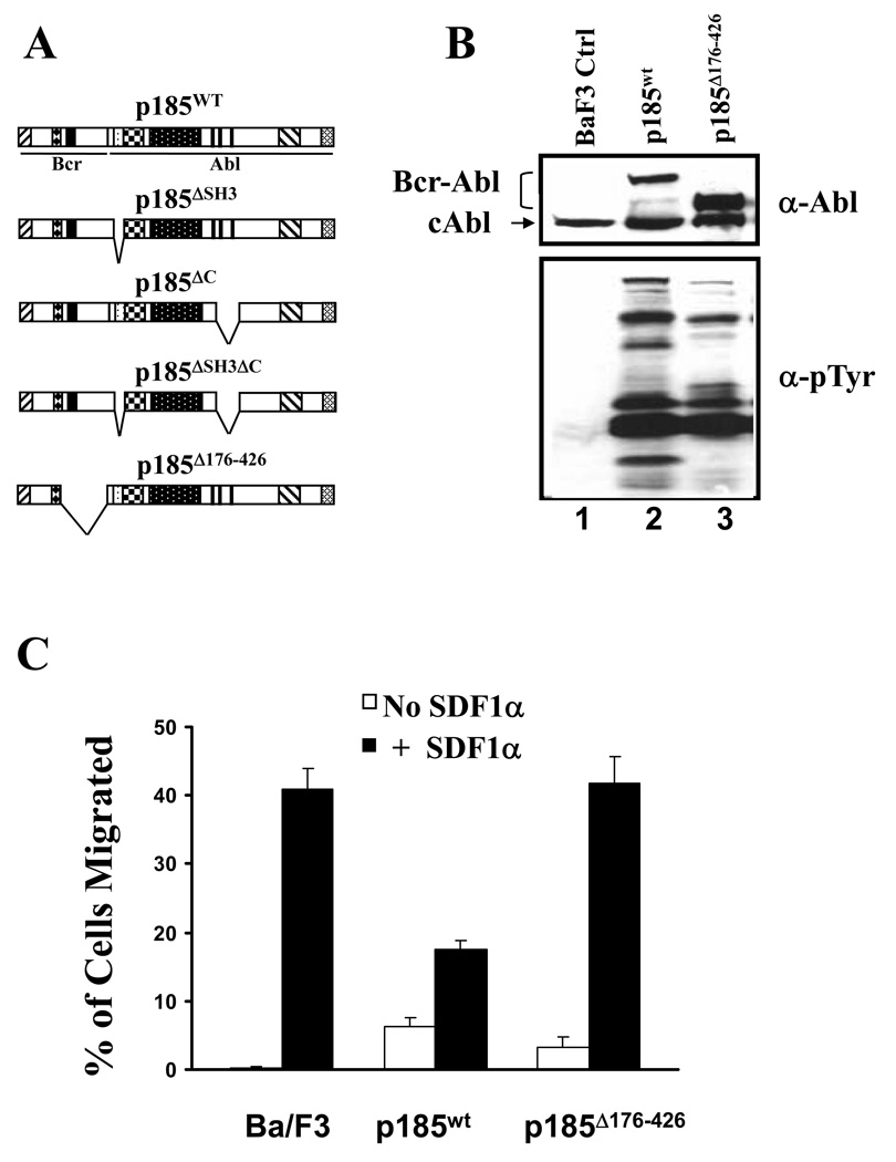
Figure 2. A mutant Bcr-Abl with deletion in Bcr sequences failed to inhibit SDF1α-stimulated chemotaxis.
A. Schematic representation of the wild type (p185wt) and mutant forms of p185Bcr-Abl. B. Profiles of protein tyrosine phosphorylation in Ba/F3 cells transduced with control retrovirus (BaF3 Ctrl) or retroviruses expressing either p185wt or p185Δ176–426, as indicated. The cells were starved in RPMI-1640 medium containing 0.1% bovine serum albumin for 6 hours. Total lysates from 1×10⁶ cells were subjected to Western blot analysis with anti-Abl antibody (α-Abl) and anti-phosphotyrosine (α-Tyr) antibodies. The positions of the wild type and mutant form of p185Bcr-Abl as well as endogenous c-Abl are indicated. C. The p185Δ176–426 failed to inhibit SDF1α-stimulated chemotaxis in Ba/F3 cells. The data represents one of three independent cell migration experiments. The vertical axis shows the percentage of migrated cells and is expressed as the average +/− S.D. calculated from triplicate wells.

