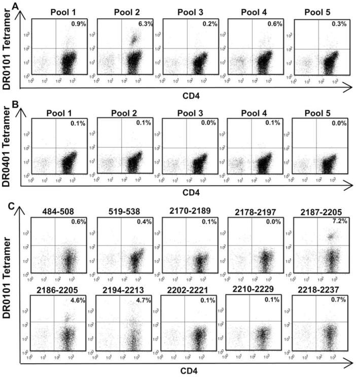
Figure 2. T-cell epitope mapping for haemophilic subject IV-2.
CD4+ T cells were stimulated with pooled peptides spanning the FVIII C2 domain sequence. Eighteen days later, the cells were incubated with PE-labeled DR0101 tetramers loaded with FVIII C2 peptide pools (A) or with DR0401 tetramers loaded with FVIII C2 peptide pools (B) and antibodies as described in Methods. Decoding of positive CD4+ responses to DR0101 tetramers loaded with peptide pools 1 and 2 was carried out 22 days after stimulation of total CD4+ cells (top row) or CD4+CD25+-depleted CD4+ cells (bottom row), respectively (C). Decoding of DR0101-restricted responses to peptide pool 1 using tetramers loaded with individual peptides comprising the pool is shown in the top row. Decoding of DR0101-restricted responses to peptide pool 2 is shown in the bottom row.

