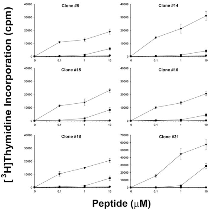
Figure 5. Antigen-specific proliferation of T-cell clones from haemophilic subject IV-2.

Resting T-cell clones #5, 14, 15, 16, 18, and 21 were stimulated with PBMCs from a healthy DRB1*0101 donor plus wild-type peptide FVIII2194-2213 (triangle symbols), haemophilic peptide FVIII2194-2213 2201P (square symbols), or irrelevant peptide FVIII519-538 (circle symbols) at 0, 0.1, 1.0, and 10 μM final concentration. [3H]thymidine uptake was measured. Data show mean ± SD of triplicate determinations.
