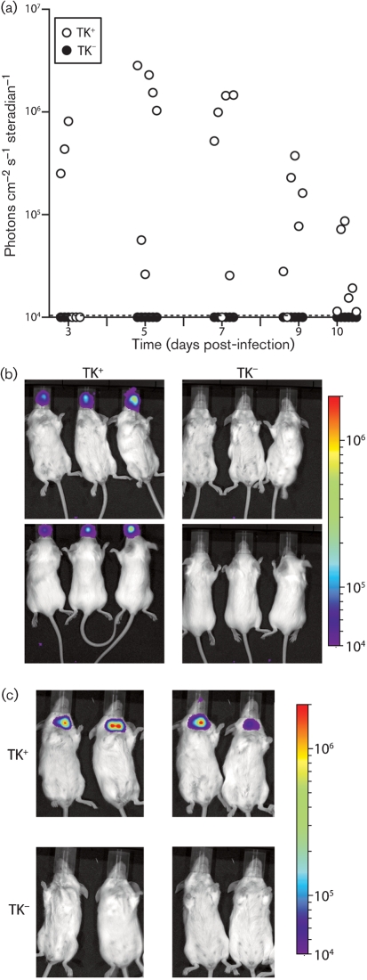
Fig. 2.
TK− MuHV-4 infection of the upper respiratory tract. (a) Mice were inoculated intranasally with TK+ or TK− luciferase+ MuHV-4 (103 p.f.u. in 5 μl), then imaged for luciferase expression over the next 10 days. Each point shows the maximum radiance signal of one mouse. The dashed line shows the lower limit of assay sensitivity. (b) Example dorsal and ventral views of luciferase signals 3 days after infection with 5 μl TK+ or TK− luciferase+ MuHV-4. (c) Luciferase signals at 10 days after infection of four mice each with 5 μl TK+ or TK− luciferase+ MuHV-4. The neck signal is from the superficial cervical lymph nodes.

