Fig. 3.
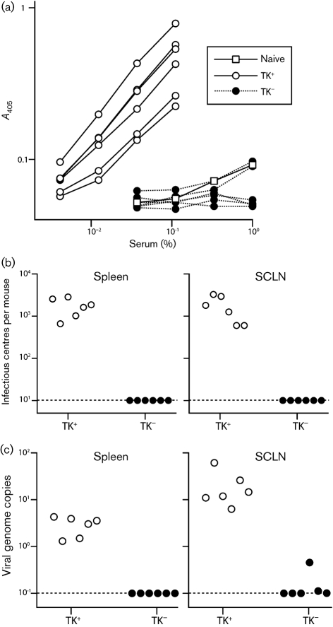
No late colonization of mice by TK− MuHV-4 delivered to the upper respiratory tract. (a) Mice were inoculated intranasally (103 p.f.u., 5 μl) with TK+ or TK− luciferase+ MuHV-4, then analysed for MuHV-4-specific IgG at 1 month post-infection by ELISA. Each line shows the result for one mouse. Pooled naive sera provide the negative control. (b) The same mice as in (a) were analysed for reactivatable splenic virus by infectious centre assay. Each point shows the titre for one mouse. The dashed lines show lower limits of assay sensitivity. (c) The same samples as in (b) were analysed for viral genomes by real-time PCR of DNA from spleens or SCLN. Each point shows the viral genome copy number of one mouse, normalized by the host genome copy number of the same sample (1000× viral genomes per host genome). The dashed lines show lower limits of assay sensitivity.

