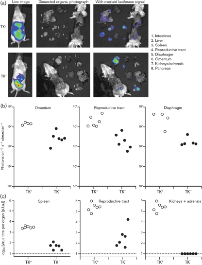
Fig. 6.
Intraperitoneal infection by TK− MuHV-4: individual organs. (a) Mice were infected intraperitoneally with TK+ or TK− luciferase+ MuHV-4 (103 p.f.u.), and 5 days later imaged for luciferase expression, first live and then post-mortem after dissection. Representative examples are shown. (b) Quantification of data equivalent to those of (a), showing maximum radiance values for the omentum, reproductive tract and diaphragm of six mice per group. Each point shows the signal for one mouse. The dashed lines show signal detection limits. (c) Virus titres of samples from equivalent infections to (b), measured by plaque assay. Each point shows the titre for one mouse. The dashed lines show lower limits of detection.

