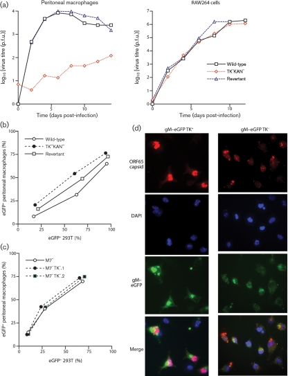
Fig. 7.
Macrophage infection by TK+ and TK− MuHV-4. (a) Peritoneal macrophages from naive mice or RAW-264 cells were infected (0.01 p.f.u. per cell, 2 h) with TK+ (wild-type or revertant) or TK− viruses, then washed twice in PBS and cultured at 37 °C. Samples were titrated for infectious virus by plaque assay at the times shown. (b) 293T epithelial cells or peritoneal macrophages were exposed to eGFP+ TK+ or TK− viruses at different multiplicities, then 24 h later assayed for eGFP expression by flow cytometry. Since MuHV-4 infects 293T cells better than peritoneal macrophages, the infecting doses for the latter were 10 times higher for each virus. The peritoneal macrophages were treated with LPS (100 ng ml−1, 6 h) before assay to maximize HCMV IE1 promoter-driven viral eGFP expression. (c) As in (b), we compared TK+ and TK− viruses for their capacity to infect either 293T cells or peritoneal macrophages, except that all viruses were additionally disrupted for M7 (gp150). This allowed us to use equivalent virus doses for each cell type, as M7− MuHV-4 infects macrophages more efficiently than the wild-type. (d) Peritoneal macrophages were exposed (2 p.f.u. per cell) to TK+ or TK− viruses carrying eGFP-tagged gM (green). The cells were fixed 18 h later, permeabilized and stained for the ORF65 capsid component (red). Nuclei were counterstained with DAPI (blue).

