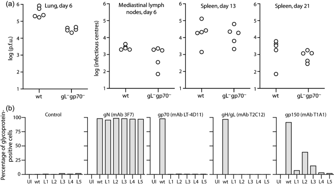
Fig. 4.
In vivo infection with gL−gp70− MuHV-4. (a) Mice were infected intranasally (300 p.f.u.) with wt or gL−gp70− MuHV-4, then analysed for infectious virus in lungs by plaque assay and for latent virus in lymphoid tissue by infectious centre assay. Each point shows the titre of one mouse. gL−gp70− titres were significantly lower than wt in lungs at day 6 (P<0.001 by Student's t-test) but not in lymphoid tissue. (b) gL−gp70− viruses from the individual mouse lungs in (a) (L1-L5) were propagated in BHK-21 cells for 7 days then analysed for glycoprotein expression by flow cytometry. Cells were scored as stained or not, based on a gate excluding >99 % of uninfected cells. Uninfected (UI) and wt virus-infected (wt) BHK-21 cells provided controls. Each bar shows 2×10⁴ cells. For equivalent gN expression, gp150 expression by the gL−gp70− knockouts was significantly reduced compared with wt (P<10−5 by χ2 test).

