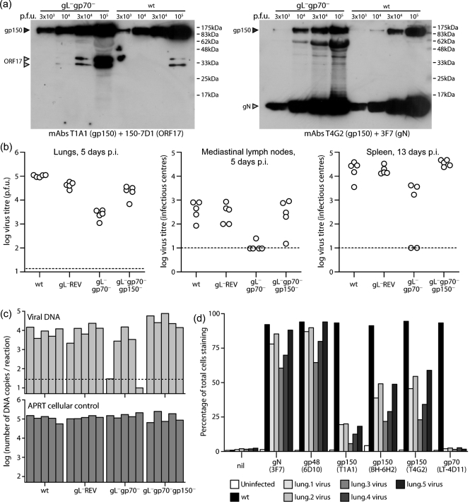
Fig. 6.
In vivo infection with gL−gp70−gp150+ MuHV-4. (a) The gL−gp70−.2 mutant was analysed for gp150 content by immunoblot with mAbs that recognize gp150 residues 152–269 (T1A1) or 108–151 (T4G2) (Gillet et al., 2007d). gN and the ORF17 capsid component provided loading controls. The 62 kDa band in the T4G2+3F7 immunoblot of the gL−gp70− stock is non-specific staining of bovine albumin. Because gL−gp70− stocks had lower titres than wt, more albumin was present at comparable infectivity. (b) Mice were infected intranasally (300 p.f.u.) with wt or gL−gp70− viruses analysed in (a), then titrated for infectious virus in lungs by plaque assay and for latent virus in lymphoid tissue by infectious centre assay. Each point shows the titre of one mouse. The horizontal dashed lines show lower limits of detection. gL−gp70−, but not gL−gp70−gp150− virus titres were significantly lower than wt in all sites (P<0.01 by Student's t-test). (c) DNA was extracted from the spleens in (b) and viral genome copy numbers determined by real-time PCR. Each bar shows the average of triplicate reactions. The horizontal dashed line shows the average of three uninfected controls. (d) gL−gp70− viruses were recovered from infected lungs by propagation in BHK-21 cells for 14 days, then analysed for virion glycoprotein expression by flow cytometry. BHK-21 cells either uninfected or infected overnight with wt MuHV-4 (1 p.f.u. cell−1, 18 h), provided staining controls. nil, Secondary antibody only. Each bar shows 2×10⁴ cells. mAbs T4G2, T1A1 and BH-6H2 recognize different gp150 epitopes.

