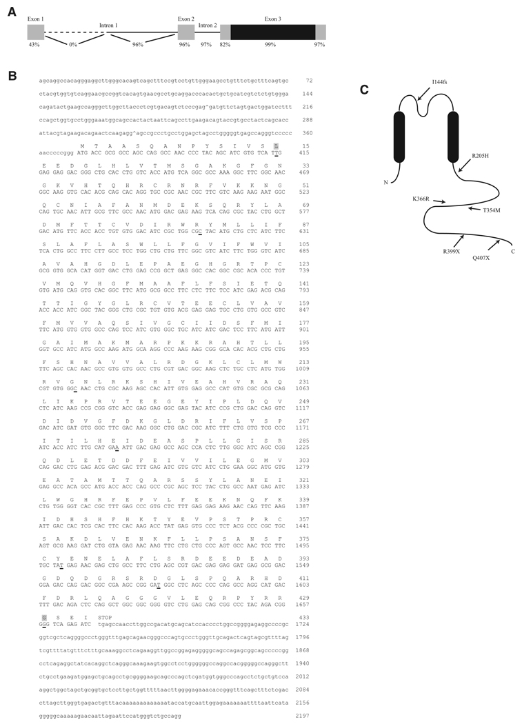
Figure 1. Structure and Sequence of KCNJ18 and Kir2.6.
(A) KCNJ18 shares a high degree of identity with KCNJ12 in both exons (boxes) and introns (lines). The coding region of both KCNJ18 and KCNJ12 is contained within exon 3 (black region). The first intron of KCNJ12 is longer than that of KCNJ18, causing 0% identity in this nonoverlapping region (dotted line).
(B) KCNJ18 sequence-exon boundaries are denoted with a caret (^). Coding sequence is capitalized with the corresponding amino acid above. Underlined nucleotides denote differences between KCNJ18 and KCNJ12, with nonsynonymous differences having a gray background.
(C) Diagram of Kir2.6 with the relative locations of TPP associated mutations.
See also Figure S1.

