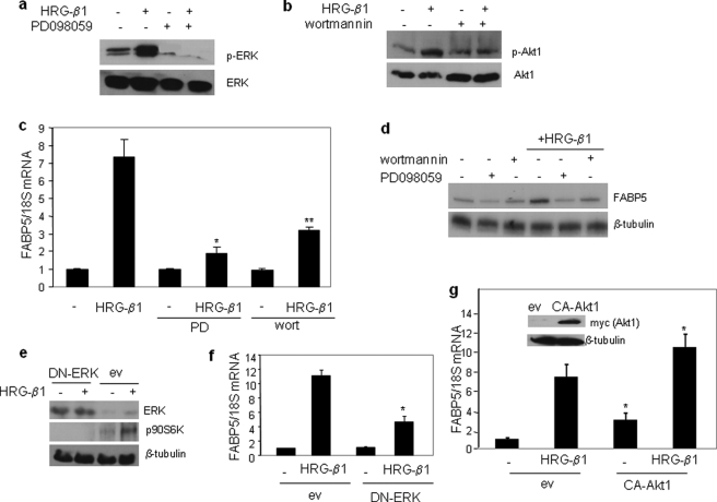
FIGURE 2.
Up-regulation of FABP5 by HRG-β1 is mediated by the ERK and PI3K pathways. a, serum-starved MCF-7 cells were treated with the MEK inhibitor PDO98059 (20 μm) for 1 h before the addition of HRG-β1 (30 ng/ml, 15 min). Cell lysates were analyzed by immunoblots using antibodies against phospho-ERK (p-ERK) and total ERK. b, serum-starved MCF-7 cells were treated with the PI3K inhibitor wortmannin (100 nm) for 1 h before the addition of HRG-β1 (30 ng/ml, 15 min). Cell lysates were analyzed by immunoblots using antibodies against phospho-Akt (p-Akt) and total Akt. c, serum-starved MCF-7 cells were treated with PDO98059 (PD, 20 μm) or wortmannin (100 nm) for 1 h before the addition of HRG-β1 (30 ng/ml, 4 h). FABP5 mRNA levels were measured by Q-PCR. Data are the mean ± S.D. (n = 3). p = 0.006 (*) and p = 0.014 (**) versus HRG-β1 alone. d, serum-starved MCF-7 cells were treated with PDO98059 (20 μm) or wortmannin (100 nm) for 1 h before the addition of HRG-β1 (30 ng/ml, 24 h). Cell lysates were analyzed by immunoblots. e, MCF-7 cells were transfected with an empty vector (ev) or an expression vector for the dominant negative ERK-K52R (DN-ERK), serum-starved overnight, and then treated with HRG-β1 (15 min.). Cell lysates were analyzed by immunoblots using antibodies recognizing ERK or its substrate p-p90S6K. β-Tubulin was used as a loading control. f, MCF-7 cells were transfected with denoted plasmids, serum-starved overnight, and then treated with HRG-β1 (4 h). Expression of FABP5 mRNA was measured by Q-PCR. Data are the mean ± S.D. (n = 3). *, p = 0.018 versus HRG-β1-treated cells transfected with an empty vector. g, MCF-7 cells were transfected with a control vector or a vector harboring Myc-tagged constitutively active (CA)-myristoylated Akt1. 36 h later cells were serum-starved overnight and treated with HRG-β1 (30 ng/ml, 4 h). FABP5 mRNA levels were measured by Q-PCR. Data are the mean ± S.D. (n = 3). *, p < 0.03 versus respective empty vector-transfected controls. Inset, shown are immunoblots demonstrating expression of myc-CA-Akt1.

