FIGURE 3.
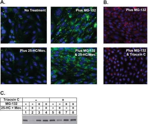
Proteasome inhibition stimulates formation of lipid droplets in CHO-K1 cells. A, on day 0 CHO-K1 cells were set up on glass coverslips at a density of 2 × 10⁵ cells per well of 6-well plates in medium A containing 5% FCS. On day 1 cells were refed medium A supplemented with 5% LPDS, 10 μm compactin, and 50 μm mevalonate (Mev.). After 16 h at 37 °C, cells were switched to medium A containing 5% LPDS and 10 μm compactin in the absence or presence of 10 μm MG-132 and 1 μg/ml 25-hydroxycholesterol plus 10 mm mevalonate as indicated. After incubation for 5 h at 37 °C, cells were washed twice with PBS, fixed with 4% paraformaldehyde, permeabilized with 0.1% Triton X-100, and stained with 10 μg/ml BODIPY 493/503 (green) to visualize lipid droplets or Hoechst reagent (blue) to visualize nuclei using standard procedures. Images were acquired with Zeiss AxioPlan 2E (Thornwood, NY) and a Hamamatsu monochrome camera (63× objective, Bridgewater, NJ). B and C, on day 0 CHO-K1 cells were plated at 2 × 10⁵ cells per well of 6-well plates with glass coverslips or at 4 × 10⁵ cells per 100 mm dish in medium A containing 5% FCS. On day 2 cells were refed medium A supplemented with 5% delipidated fetal calf serum, 10 μm compactin, and 50 μm mevalonate. After 16 h at 37 °C, cells were switched to medium A containing 5% LPDS and 10 μm compactin without (−) or with (+) 9.6 μm triacsin C. After 3.25 h at 37 °C, cells were treated in the absence (−) or presence (+) of 9.6 μm triacsin C, 10 μm MG-132, and 1 μg/ml 25-HC plus 10 mm mevalonate as indicated. B, after 4 h at 37 °C cells on the coverslips were stained with Oil Red O and Hoechst by standard methods. C, cells on the 100-mm dishes were harvested after 4 h at 37 °C, and membrane fractions were obtained as described under “Experimental Procedures.” Aliquots of the membrane fractions (10 μg protein/lane) were subjected to SDS-PAGE and transferred to supported nitrocellulose membranes followed by immunoblot analysis with 5 μg/ml monoclonal IgG-A9 (against reductase).

