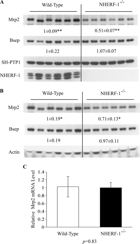
FIGURE 5.
Expression of Mrp2 protein, but not mRNA, is reduced in liver lysates and membranes from NHERF-1−/− mice. A, representative immunoblots of whole cell lysates from wild-type (n = 6) and NHERF-1−/− (n = 6) mouse liver demonstrate reduction in Mrp2 but not Bsep protein in NHERF-1−/− mice. B, representative immunoblots of membrane-enriched fractions confirm these findings. In both A and B, numbers represent the relative ratio of the respective protein bands in wild-type and NHERF-1−/− mice by densitometry analysis. Data are normalized to SH-PTP1 in whole cell lysates (A) and to actin in membrane-enriched fractions (B). The amount of protein from the wild-type is set as 1. Values represent the means ± S.D. of six individual animals in each group. ** indicates p < 1 × 10⁻⁶. * indicates p < 0.05. C, relative levels of Mrp2 mRNA in the liver of wild-type (n = 6) and NHERF-1−/− (n = 6) mice quantified by real-time PCR. Data are normalized to Gapdh, and the amount of Mrp2 mRNA from the wild-type is set as 1. Values represent the means ± S.D. of six individual animals in each group.

