FIGURE 6.
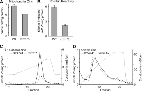
Mitochondrial Zn defect in mzm1Δ cells. Mitochondria were isolated for both WT and mzm1Δ from cells grown in yeast peptone 1% dextrose. Panel A, metal analysis of mitochondria (200 μg; n = 3). Panel B, mitochondria (50 μg) were sonicated in 0.1 ml 10 mm Tris, and the clarified lysate incubated with Rhodzin-3 to asses Zn-dependent fluorescence. Following fluorescent measurements, lysates were analyzed for metal content by ICP-OES (n = 3). Panel C, MonoS fractionation of mzm1Δ soluble mitochondrial lysate. Panel D, MonoQ fractionation of mzm1Δ soluble mitochondrial lysate.

