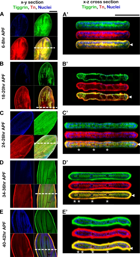
FIGURE 2.
Tiggrin and O-glycans show dynamic and specific localization during pupal wing development. Shown are stages of wild type pupal wing development 6–8 h after puparium formation (6–8 h APF), 18–20 h after puparium formation (18–20 h APF), 24–26 h after puparium formation (24–26 h APF), 34–36 h after puparium formation (34–36 h APF), and 40–42 h after puparium formation (40–42 h APF). Wings were stained for Tiggrin (green), O-glycans (red), and DAPI (blue). Merged images show Tiggrin and O-glycan co-staining in yellow. Images shown are X-Y sections (A–E) or X-Z optical cross-sections (A′, B′, C′, D′, and E′). White dashed lines indicate regions used to produce X-Z optical cross-sections. Arrowheads at the right denote the basal dorsal/ventral cell layer interface, and asterisks denote the position of wing veins. Black bar, 100 μm.

