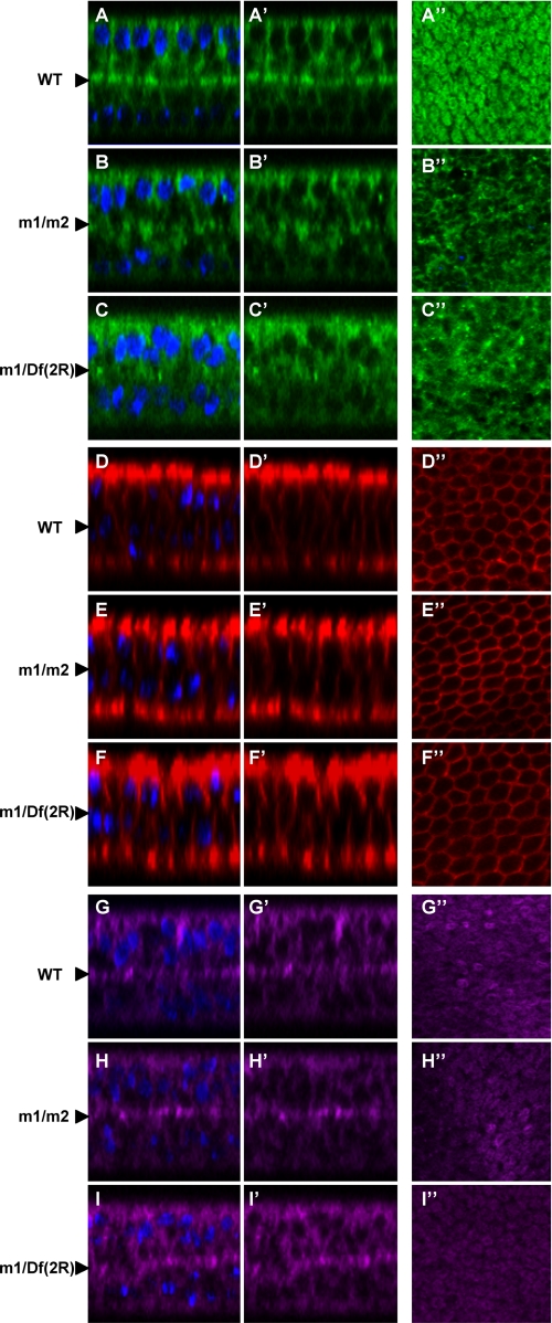
FIGURE 4.
Localization of Tiggrin, but not other proteins, is affected in pgant3 mutant pupal wings. Wild type (WT) (A–A″, D–D″, and G–G″), pgant3m1/pgant3m2 (m1/m2) (B–B″, E–E″, and H–H″), and pgant3m1/Df(2R)Exel6283 (m1/Df(2R)) (C–C″, F–F″, and I–I″) pupal wings at 36 h APF were stained with Tiggrin (green) (A–C″), Fasciclin III (red) (D–F″), and DE-cadherin (purple) (G–H″). Shown are X-Z optical cross-sections of pupal wings with (A–I) or without (A′–I′) DAPI staining of nuclei. Images are oriented so that dorsal is at the top and ventral is at the bottom. Arrowheads at the left denote the basal dorsal/ventral cell layer interface. X-Y confocal images of the basal region comprising the dorsal and ventral boundary (A″–I″) are also shown. In wild type wings 36 h APF, Tiggrin localizes to the dorsal/ventral cell layer interface, where cell adhesion occurs. Mutations in pgant3 result in loss of Tiggrin localization at this interface, although DE-cadherin and Fasciclin III localizations are unaffected.

