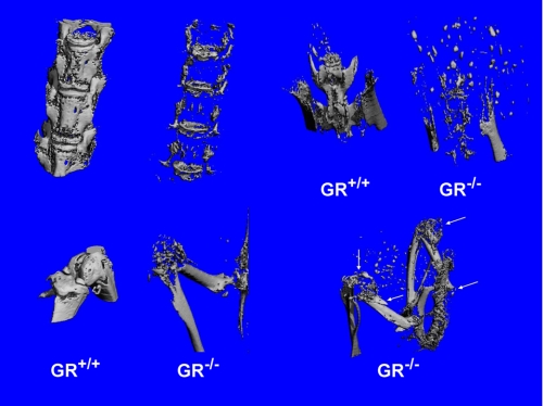
FIGURE 3.
Microcomputed tomography reconstructions of the spinal column, pelvis, knee, femur, and tibia of a WT and an affected GRKO (189-day-old female). Carcasses were scanned at 36 μm in a Scanco μCT 40 with a threshold value of 240. Note the mineral disappearance in the GRKO bones as compared with the WT and dissolution of the patella and numerous fractures (arrows) in the long bones.

