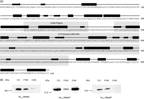
FIGURE 4.
Secondary structure predictions and localization of His10-WbbM. A, sequence and predicted secondary structure of WbbM outlining β-sheets (black arrows) and α-helices (black boxes). The GT-8 region (residues 284–549) is identified, as is a putative linked region that may separate two independent domains (WbbMN and WbbMC). B, localization of His10-WbbM (expressed from pWQ522 in E. coli CWG286), His10-WbbMN (pWQ530), and His10-WbbMC (pWQ531). Cell-free lysates (S12) were separated into soluble (S100) and membrane-containing (P100) fractions and the His-tagged proteins were detected in Western immunoblots using mouse anti-His5 monoclonal antibody followed by goat anti-mouse horseradish peroxidase-conjugated antibody. Loading was standardized to the original cell suspension (prior to lysis), allowing direct visual assessment of the cellular distribution of His10-WbbM.

