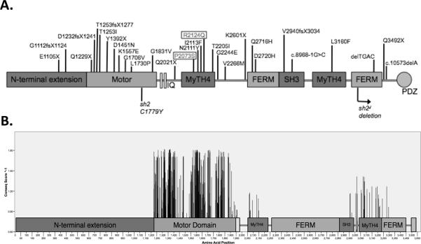
Figure 1.
Domains of the myosin XVa protein. A. Diagram showing the location of human (top) and mouse (bottom) MYO15A mutations. The p.R2124Q and p.P2073S mutations are boxed Figure is not to scale. B. Diagram showing the amino acid positions in each domain of myosin XVa (x-axis) and Conseq conservation scores for each residue (y-axis). Any residue with a Conseq score less than 7 (E-value >0) was excluded. Figure is to scale.

