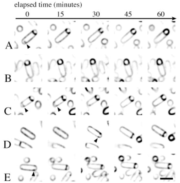
Fig. 3.
Observation of engulfment by time-lapse microscopy. Sporulation of the wild-type strain PY79 was induced by resuspension in the presence of FM 4-64 and affixed to a coverslip (Experimental procedures). The slide was heated to approximately 30°C, and five images were collected every 15 min (five columns, left to right). For ease of presentation, five individual sporangia (rows A–E) were selected from the field. Partial septa are visible within the mother cells of the sporangia shown in (A) and (C) (arrows). The bar equals 2 μm.

