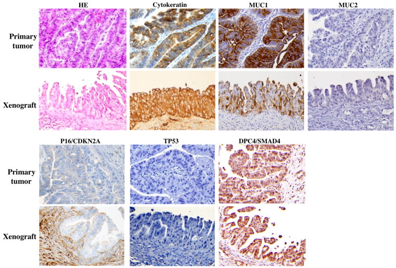
Figure 1.
Histology and immunohistochemistry of matched primary tumor and corresponding xenograft for case 1. Hematoxylin and eosin (HE) and immunohistochemical labeling for cytokeratin, MUC1, MUC2, P16/cdkn2a, TP53, and DPC4/SMAD4. Negative region of P16/cdkn2a is shown for the primary tumor, however the staining is heterogeneous as shown in supplemental figure 2. Magnification as indicated.

