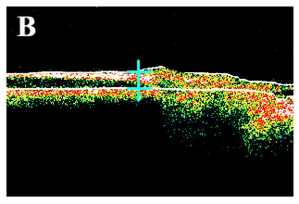
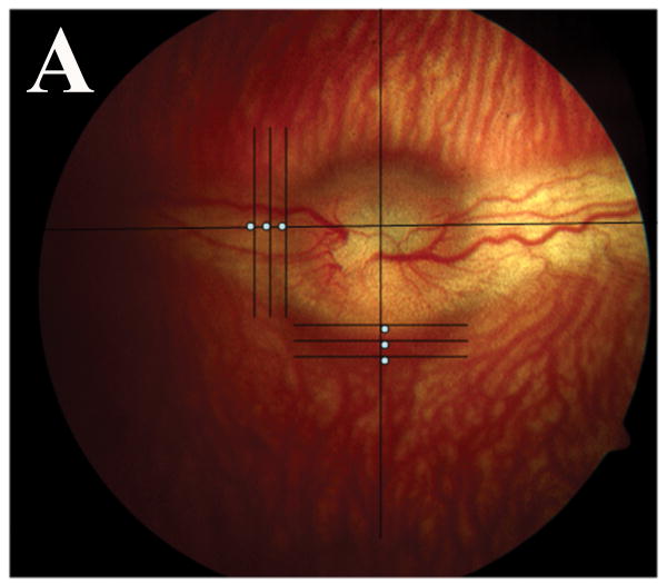
Fig. 1.

Color fundus photograph and ocular coherence tomography (OCT) of a representative rabbit. (A) Different locations at which retinal thickness was measured by OCT. Note 3 white spots in the temporal medullary wing and below the optic disc, which are located 100 μm apart. (B) Sample retinal thickness measurement below the optic disc by OCT in a representative rabbit. The scan is along a vertical line at the middle inferior edge of the optic disc.

