
Figure 2. Structural analysis of nucleosomes reconstituted with H3K56E.
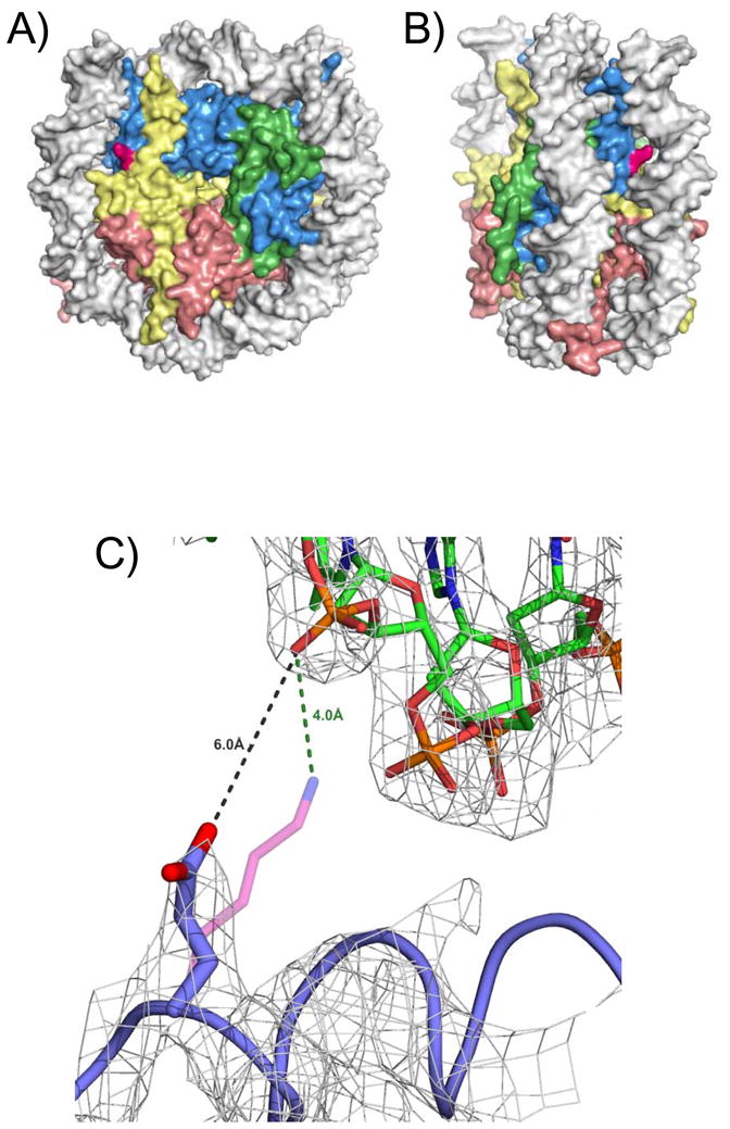
A) Location of H3K56 in the nucleosome. H3 is shown in blue, H4 in green, H2A in yellow, and H2B in red. H3K56 is indicated in magenta. The nucleosome is viewed down the superhelical axis. B) as A), but rotated around the y-axis by about 75 degrees. C) Detailed view of the structure of a nucleosome with H3K56E, superimposed onto the wild type structure. Distances between the side chains and DNA are indicated. Electron density (2Fo-Fc) is contoured at 1 sigma.