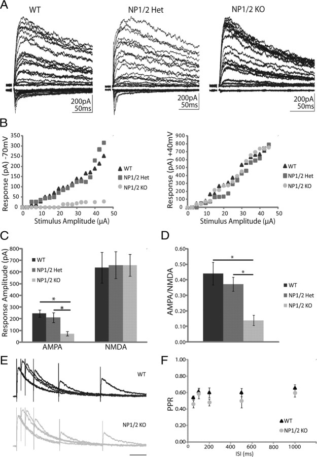
Figure 1.
NP1/2 knock-out neurons have a specific deficit in AMPAR-mediated synaptic transmission during early postnatal development (P6–P9). A, Example traces of postsynaptic responses from WT, NP1/2 Het, and NP1/2 KO dLGN neurons voltage clamped at −70 mV (downward currents) and +40 mV (upward currents) where the optic tract was stimulated over a range of stimulus intensities. B, Example input–output curves showing EPSC amplitude as a function of stimulus intensity at −70 mV and +40 mV. C, Average amplitudes of maximal AMPAR and NMDAR-mediated currents. D, Average AMPA/NMDA ratios of maximal currents. C, D, n = 9 WT animals (26 cells), 6 NP1/2 Het animals (15 cells), and 6 NP1/2 KO animals (26 cells). Data displayed as mean ± SEM and compared by one-way ANOVA followed by Bonferroni post-tests. *p < 0.05. E, Example traces from a WT neuron (upper) and a NP1/2 KO neuron (lower) voltage clamped at +40 mV and stimulated with pairs of pulses at 5 different interstimulus intervals (50, 100, 200, 500, and 1000 ms). F, The average PPRs for WTs (3 animals, 8 cells) and NP1/2 KO neurons (2 animals, 4 cells) over the 5 ISIs tested. No differences were found by two-way ANOVA followed by Bonferroni post-tests.

