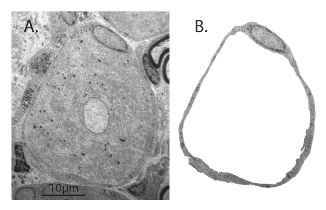
Figure 7.

Electron microscopy image of rat dorsal root ganglion. A. The center of the image shows the soma of a sensory neuron of the small-dark category. B. An envelope of satellite glial cells, including the nucleus of one, surrounds the neuronal soma (removed from the image).
