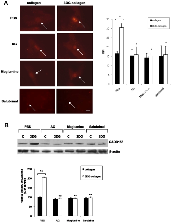
Figure 2. Effect of ER stress inhibitor salubrinal on 3DG-collagen-induced GADD153 expression.
A, Fibroblasts were cultured in chamber slides coated with native collagen or 1 mM 3DG-collagen with or without 5 mM AG or 40 mM meglumine for 24 h. Also, fibroblasts were pretreated for 1 h with or without 40 µM salubrinal and then cultured on native collagen or 3DG-collagen for 24 h. Fibroblasts were stained and analyzed for expression of GADD153 in the nucleus by immunofluorescence analysis using Cy3-conjugated secondary antibody. Mean fluorescence intensity (MFI) of GADD153 in the nucleus was measured using ImageJ from ten representative fibroblasts. Images were taken at 40× magnification on an epi-fluorescent microscope. Arrows indicate nuclei containing GADD153. The bars represent the MFI values from each experimental condition. Scale bar represents 10 µm. B, Fibroblasts were treated as in A followed by Western blot for GADD153 expression. β-actin was used as a loading control. The bars represent the densitometric value for each experimental condition. All comparisons are made against 3DG-collagen treated with PBS unless otherwise indicated. Data are mean±SD (n = 3), **P<0.0005, *P<0.007.

