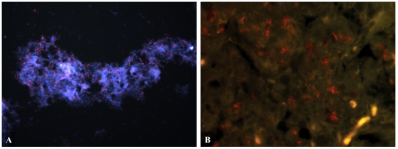
Figure 2. FISH detecting Mtb in culture and mouse lung tissue.
Mtb (red) was detected in (A) hypoxic culture and (B) GKO mouse lung tissue at 4 weeks post infection. Mice were exposed to a low-dose aerosol infection (LDA) with M. tuberculosis strain Erdman (TMCC 107). 5 µm formalin fixed paraffin embedded sections were dewaxed and rehydrated. Three fluorescent Alexafluor 568 nm labeled ssDNA probes designed to detect the 16 s rRNA subunit were hybridized overnight at 37°C after digesting sections for 1 hr with lysozyme at 10 µg/ml. Sections were counterstained with DAPI and mounted. Pictures were taken using FITC and TRITC filters at (A) 400× magnification and (B) 1000× magnification.

