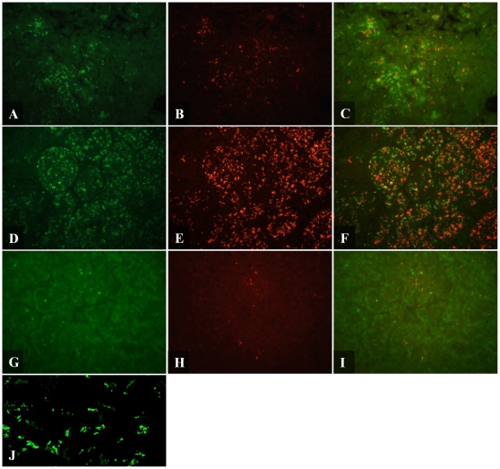
Figure 3. Fluorescent micrograph of Mtb detected by IF and AR in mouse and guinea pig.
Mtb was detected by IF (A,D,G,J), AR (B,E,H) and combined IF-AR (C,F,I) in C57BL/6 mouse lung (A–C), GKO mouse lung (D–F) and guinea pig lung (G–I). 5 µm formalin fixed paraffin embedded sections were dewaxed, rehydrated and detected by IF. Rabbit polyclonal antibody against whole Mtb cell lysate was applied overnight at 4°C and detected using an Alexafluor 488 labeled secondary antibody (green). Slides were then stained with AR (orange/red) for 30 mins then destained. Pictures were taken using a FITC and TRITC filter at 400× magnification. (J) Confocal micrograph of Mtb detected by IF in GKO lung tissue at 1000× magnification with 1.6× digital zoom.

