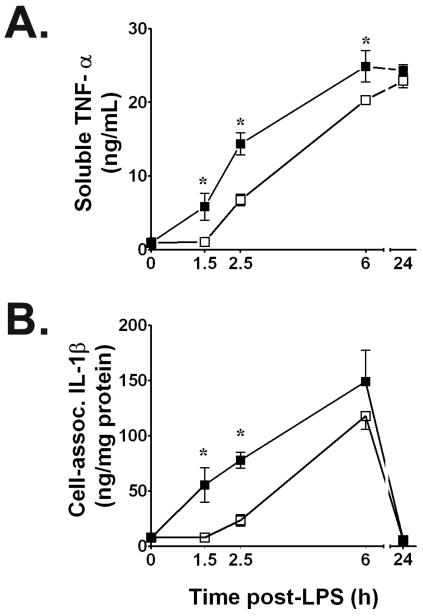
Figure 1.
A. Brief hypoxia significantly suppresses soluble immunoreactive TNF-α secretion induced by E. coli serotype O55:B5 LPS (100 ng/mL) into supernatants of NR8383 rat alveolar macrophages. B. Time course of IL-1β production in E. coli LPS-stimulated cell lysates assayed by IL-1β-specific ELISA for total protein at time points shown after adding 100 ng LPS/mL. Values in both panels are means ± SD of quadruplicate determinations from at least three experiments per time point. Supernatants contained only trace amounts of IL-1β protein, while cell lysates contained only trace amounts of immunoreactive TNF-α protein. Open squares, LPS + hypoxia or LPS + combined hypoxia/reoxygenation (H/R) time-matched values. Filled squares, normoxic LPS control values; * P < 0.01 vs. time-matched normoxic LPS control value.

