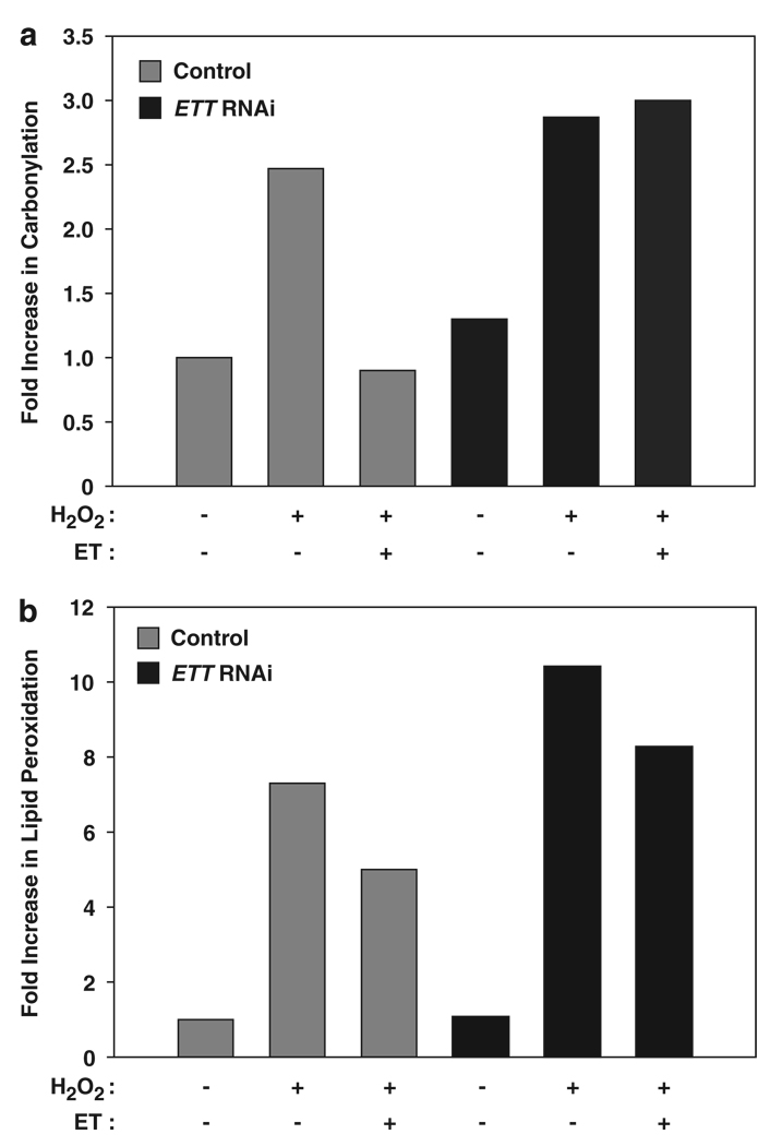
Figure 3.
Depletion of the ET transporter leads to increased damage to cellular proteins and lipids. (a) Control- and ETT-depleted HeLa cells were treated with 0.5 mM H2O2 or 16 h after preincubation with 1 mM ET and assayed for protein oxidation using 2,4-dinitrophenylhydrazine (DNPH), which reacts with protein carbonyl groups generated by oxidation. The DNPH-derivatized samples were electrophoresed and subjected to western blotting using anti-DNPH antibodies. The signals were quantified using densitometry and expressed as a fold increase in protein carbonylation compared with the untreated control. The data shown are from a representative experiment repeated at least three times. The ETT-depleted cells (black bars) show higher levels of oxidation compared with control cells (gray bars). (b) ETT cells show a higher level of lipid peroxidation. Cells were pretreated with 1 mM ET 72 h post transfection and lipid peroxidation induced using a mixture of 30 µM FeSO4 and 100 µM H2O2 for 2 h at 37°C. The cells were then assayed using the Thiobarbituric Acid Reactive Substances (TBARS) Assay to quantify the levels of Malondialdehyde (MDA), which is a product of lipid peroxidation. The ETT-depleted cells undergo higher oxidation than control cells (Black versus gray bars). Data shown is a representative of three independent experiments

