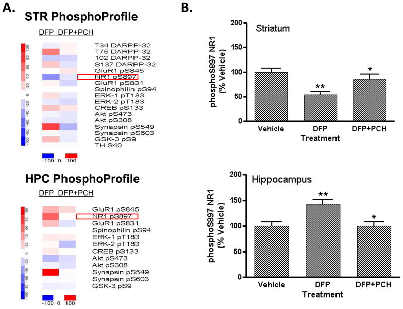
Figure 3. Effect of DFP treatment on brain protein phosphorylation in FVB mice: reversal of effects on NR1 phosphorylation by the muscarinic antagonist, PCH.
Male, adult FVB mice were injected (s.c.) with vehicle solution (50% DMSO) or PCH (25 mg/kg). Thirty min later mice received either vehicle (50% DMSO) or DFP (4 mg/kg i.p.). The mice were then sacrificed by focused microwave irradiation of the head 120 min later. Levels of each phospho-site were detected in striatal and hippocampal tissue by immunoblotting and antibody binding revealed using dual-channel fluorescent detection methods (LiCor Odyssey). Levels of immunoreactivity detected for each phospho-site were normalized for total levels of the phosphoprotein. (A) Heat map representations showing changes in phosphorylation in striatum (top) and hippocampus (bottom) of mice treated with DFP alone or DFP after PCH pre-treatment. Data were plotted using dChip 1.3 software, as described in the legend to Figure 2 above. (B) Effect of DFP and PCH pretreatment on NR1 phosphorylation in mouse striatum (top) and hippocampus (bottom). Levels of S897-phosphorylated NR1 and total NR1 were detected, revealed and quantitated, as described above. Data are expressed as a percent ± SEM of levels in the striatum or hippocampus of vehicle-treated control mice. (*p<0.01 compared with DFP alone; **p<0.001 compared with vehicle-injected controls, ANOVA with Newman-Keuls post-hoc test, n=5–6 mice/group).

