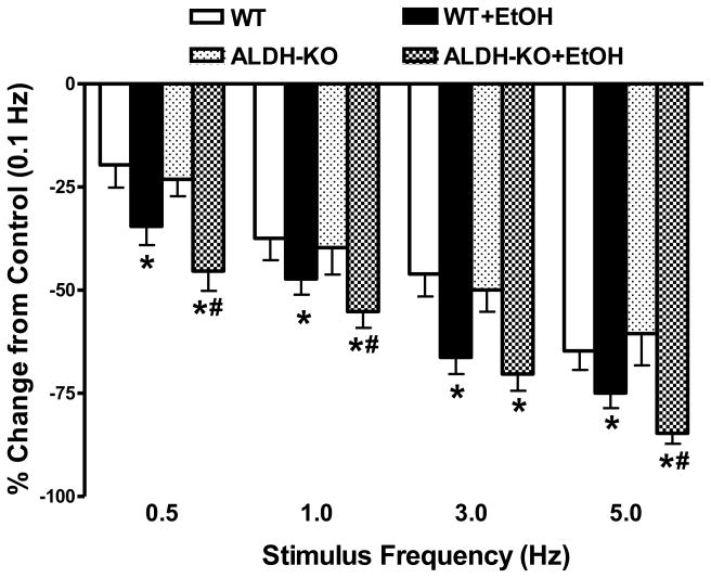
Fig. 3.
Effect of increasing stimulates frequency (0.1 – 5.0 Hz) on peak shortening amplitude in cardiomyocytes from wild-type (WT) and ALDH2 knockout (ALDH-KO) transgenic mice following acute ethanol (EtOH) exposure. Change in peak shortening amplitude was normalized to the peak shortening amplitude obtained at 0.1 Hz from the same cell. Mean ± SEM, Cell number from each group was presented in parentheses, *p < 0.05 vs. WT group; #p < 0.05 vs. WT+EtOH group.

