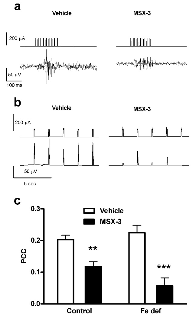
Figure 6.
A2A receptor antagonist-induced blockade of the motor output induced by cortical electrical stimulation in rats with control or iron-deficient (Fe def) diet for three weeks. (a) Representative recordings of the input signal (current pulses) delivered from the stimulator in the orofacial area of the motor cortex (upper traces) and the EMG output signal obtained from the jaw muscles (lower traces), after the administration of either saline (left traces) or the A2A receptor antagonist MSX-3 (3 mg/kg, i.p.; right traces). (b) Representative input stimulator signal power (time constant 0.01 sec; upper traces) and output electromyographic (EMG) signal power (time constant 0.01 sec; lower traces) after the administration of either saline (left traces) or MSX-3 (right traces). (c) The systemic administration of MSX-3 (1 mg/kg, i.p.) significantly decreased the power correlation coefficient (PCC) in rats with control or iron-deficient (Fe def) diet. Results are expressed as means ± S.E.M. n= 7–8 per group); **,***: significantly different compared to respective vehicle (p<0.01 and p<0.001, respectively). MSX-3 was significantly most effective in iron-deficient rats than in controls (bifactorial ANOVA: significant drug-diet interaction effect; p<0.05).

