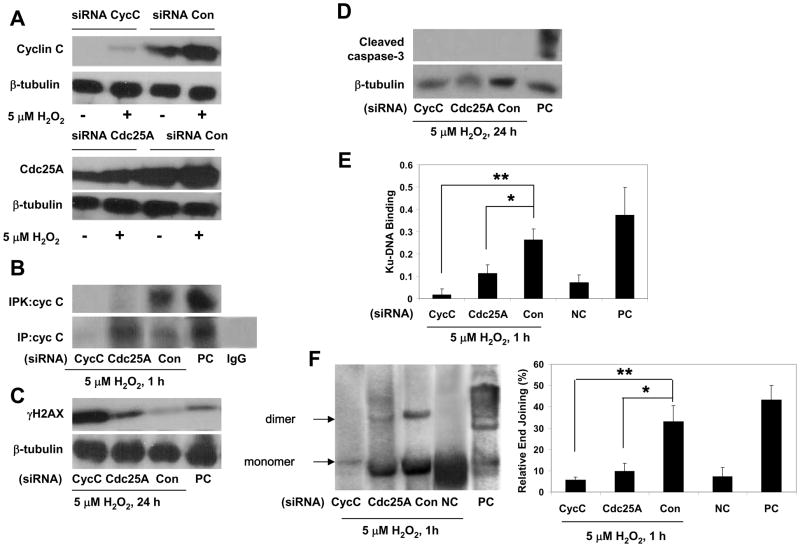
Figure 4. Blockade of cell cycle entry attenuates NHEJ activation in postmitotic neurons.
(A, B) Cortical neurons transfected either with cyclin C (cycC), Cdc25A or control (Con) siRNA were exposed to 5 μM H2O2 for the indicated durations and analyzed by immunoblotting for the expression of cyclin C and Cdc25A or by immune precipitation (IP) with anticyclin C antibody and following in vitro kinase activity assay using Rb679 as substrate (IPK). Positive control (PC), lysates from proliferating HeLa cells. Isospefic control for anticyclin C (IgG), normal rabbit IgG.
(C, D) Immunoblot analysis of DSBs (γH2AX) and apoptotic caspase-3 cleavage in neurons transfected either with cyclin C (cycC), Cdc25A or control (Con) siRNA and exposed to 5 μM H2O2 for indicated durations. Positive control (PC), extracts from staurosporine-treated apoptotic Jurkat cells.
(E) Ku-DNA binding of cortical neurons transfected with either cyclin C (cycC), Cdc25A or control (Con) siRNA and exposed to 5 μM H2O2 (1 h). Negative control (NC), untreated untransfected cultures; positive control (PC), the nuclear extract of Raji cells. The values are the means and SD (n = 4); *p < 0.01; **p < 0.001.
(F) The NHEJ analysis of cortical neurons transfected with either cyclin C (cycC), Cdc25A or control (Con) siRNA and exposed to 5 μM H2O2. Negative control (NC), untreated cultures; positive control (PC), T4 ligase treated linearized plasmid DNA (pGL3 basic/XhoI). The values are the means and SD (n = 4); *p < 0.005; **p < 0.001.

