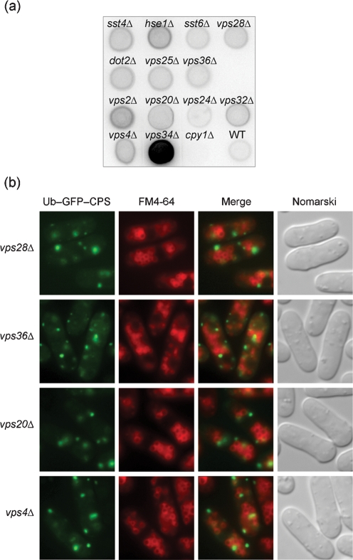
Fig. 7.
Phenotypic analyses of various class E mutants. (a) Secretion of CPY was determined using a colony blot assay. The membranes were subjected to immunoblotting with rabbit anti-CPY. Wild-type (WT), vps34Δ (positive control) and MTD2 (cpy1Δ, negative control) are included for comparison. (b) Localization of Ub–GFP–SpCPS was compared with vacuolar staining of FM4-64. Cells were processed as described in the legend to Fig. 6(a). vps28Δ, vps36Δ and vps20Δ are representative of ESCRT-I, ESCRT-II and ESCRT-III, respectively. All other mutants exhibited the same patterns of fluorescence.

