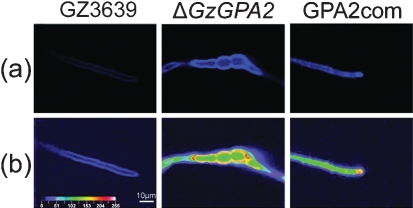
Fig. 5.
Chitin accumulation in G. zeae ΔGzGPA2 strain. GZ3639, wild-type strain; ΔGzGPA2, GzGPA2-deleted strain; GPA2com, GzGPA2-complemented strain derived from ΔGzGPA2. (a) Chitin in the mycelia of each strain was stained with a Calcofluor white solution and observed under UV (420 nm) light. (b) Quantification of fluorescence by laser scanning microscopy. The intensity scale for quantification is indicated on the bottom left, ranging from violet for a weak signal to white for strong fluorescence.

