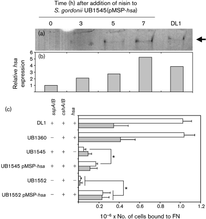
Fig. 3.
Effect of Hsa complementation on adhesion of S. gordonii to FN. The expression of Hsa from pMSP-hsa was monitored by lectin blotting (a). Cell-wall proteins were extracted from S. gordonii UB1545(pMSP-hsa) at different times following induction of hsa expression by addition of nisin to the culture. Equal amounts (20 μg) of proteins were separated by PAGE and blotted onto nitrocellulose. Membranes were probed with sWGA, which specifically recognizes Hsa, and a single band was detected at >250 kDa in each lane (arrow). Extracts from S. gordonii DL1 were included for comparison. The amount of sWGA-reactive protein in each lane was quantified by densitometry (b). (c) Adhesion of S. gordonii strains to immobilized FN was determined by staining with crystal violet, and mean values±sem from three independent experiments are shown. Significant differences between adhesion of UB1545 and UB1545(pMSP-hsa) to untreated FN, and between UB1552 and UB1552(pMSP-hsa) to untreated (white bars) or sialidase-treated (shaded bars) FN (Student's t-test, P<0.05), are indicated by asterisks.

