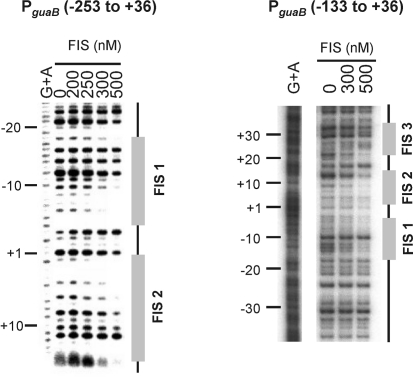
Fig. 3.
Mapping the location of FIS sites at PguaB by DNase I footprinting. A DNA fragment containing PguaB (−253 to +36) radiolabelled at the downstream end (relative to the guaB transcription start site), and a DNA fragment containing PguaB (−133 to +36) labelled at the upstream end, were employed in DNase I footprinting in the presence of different concentrations of FIS (as shown). Nucleotide positions are shown relative to the guaB transcription start site, and lanes containing the G+A ladder are indicated accordingly.

