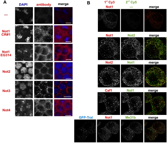
FIGURE 2.
Localization of subunits of the CCR4-NOT complex. (A) Confocal images of immunostained Drosophila embryos. Blastoderm stage embryos were incubated with the antiserum indicated on the left and a Cy3-labeled secondary antibody (middle panel). DAPI was included in the mounting medium to label the nuclei (left panel). The white bar in the merge (right panel) represents 5 μm in size. Colors in the merge are blue for the DAPI staining and red for the immunostaining. (B) Colocalization of different CCR4-NOT complex subunits in S2 cells. S2 cells were sequentially stained for two different subunits of the complex. The primary antibodies used are indicated above each image. The first secondary antibody was always Cy3-labeled (left panel); the second secondary, always Cy5-labeled (middle panel). The top panel shows a control in which the second primary antibody was omitted. No Cy5 signal was seen, confirming that the first secondary antibody had been used at saturating concentrations. The bottom panel shows sequential staining for NOT1 and Me31B in cells transiently expressing GFP-Tral for labeling of P-bodies. In the merged images (right panel), the Cy3 channel is in red, the Cy5-channel in green, and GFP in blue. The overlay of red and green becomes yellow, and the overlay of green and blue becomes turquoise.

