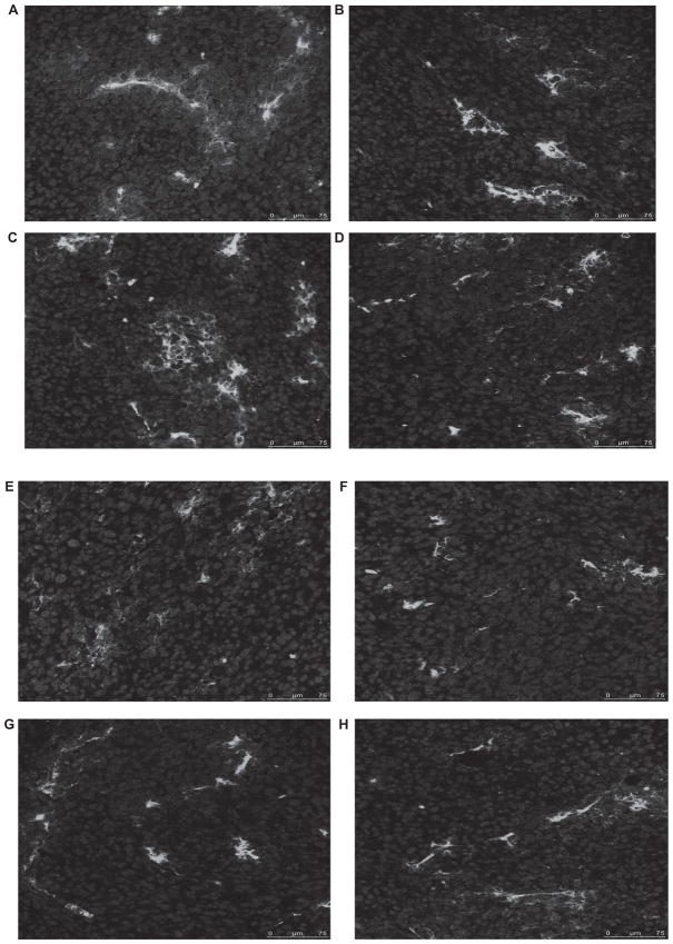
Figure 3.
Inflammatory cells infiltrate VSV23 (A, C, E, F) and control rVSV (B, D, F, G) treated tumors. A, B: CD8+ T cells. C, D: CD4+ T cells. D, e: Macrophages. F, g: neutrophils. Cohorts of N = 4, 8 to 10-week-old male BALB/c mice were injected subcutaneously on the left dorsal flank with 1 × 10⁷ JC cells. Ten days post-implantation, tumors were injected with 1 × 10⁷ pfu of VsV23, VsVsT, or VsVXn2 or vehicle alone. Viral treatments were repeated on days 3 and 5 after the initial treatment. Fourteen days after treatment was initiated, tumors were harvested, frozen, sliced into 18 μm thick sections, and treated with antibodies specificfor cell types. Images were obtained using a Leica SP5 confocal microscope at 400x magnification. Control rVSV-treated tumor images are representative of VSVST andVSVXN2 treated tumors.

