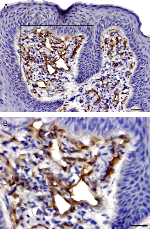
Figure 5.
Presence and distribution of CD123+ PDCs in the FP. The brown color in the sections shows the specifically stained cells. (A) CD123+ DCs exclusively present in lamina propria, with few present in epithelial compartment. The scale bar indicates 20 μm. (B) High magnification of image showing that endothelial venules in FP tissues also express CD123. The scale bar indicates 10 μm.

