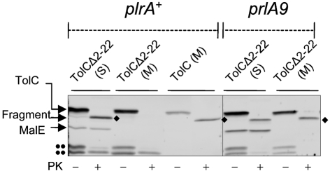
Fig. 4.
Suppression of TolCΔ2–22 by prlA9. The folding status of TolC and TolCΔ2–22 was examined in wild-type (prlA+) and S1 (prlA9) backgrounds. Membranes (M) were separated from the soluble lysates (S), as described in Fig. 1. These fractions were treated with (+) or without (–) proteinase K (PK). TolC was detected by SDS-PAGE and immunoblotting. The diamonds (⧫) indicate the 46 kDa proteinase K-resistant fragment of TolC. The double dots (••) indicate unknown TolC degradation products. Note that only the membrane fraction of cells expressing wild-type TolC was used.

