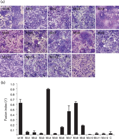
Fig. 4.
Low pH-induced syncytium formation. BSR-T7/5 cells were transfected with wt or mutant BUNV M segment cDNA constructs or left untransfected as a control (C). At 24 h post-transfection, cells were treated with low-pH medium (pH 5.3) for 5 min and syncytium formation was examined following incubation at 37 °C for a further 5 h. Cells were then stained with Giemsa solution. (a) Syncytium formation in cells transfected with wt (wt M) and mutant BUN M segment cDNAs (MΔ1–MΔ12) as indicated. The small syncytia formed by cells transfected with MΔ6 and MΔ9 cDNAs are marked by white arrows. (b) Fusion indices (f), calculated as described in Methods from cells treated with low-pH medium.

