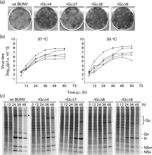
Fig. 6.
Plaque phenotypes, growth kinetics and protein synthesis profiles of wt and mutant viruses. (a) Comparison of plaque size on BHK-21 cells. Monolayers were fixed 6 days post-infection with 4 % formaldehyde and stained with Giemsa solution. (b) Virus growth curves in BHK-21 cells (at 37 and 33 °C). Cells were infected with either wt (⧫) or recombinant (□, rBUNGcΔ4; ▵, rBUNGcΔ7; ○, rBUNGcΔ8; ×, rBUNGcΔ9) viruses at an m.o.i. of 0.01 p.f.u. per cell. Virus was harvested at intervals as indicated and titrated by plaque formation in BHK-21 cells. Results are shown as the mean of two independent titrations. (c) Time-course of protein synthesis in infected BHK-21 cells. Cells were infected at an m.o.i. of 0.01 p.f.u. per cell and were labelled with 80 μCi [35S]methionine for 1 h at the time points indicated. Cell lysates were analysed on 4–12 % polyacrylamide NuPAGE gels. The positions of viral proteins are indicated; * indicates wt Gc.

