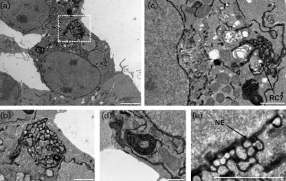
Fig. 6.
ER and nuclear envelope reorganization observed in FCV p30-expressing 293T cells. Transmission electron microscopy (TEM) of 293T cells expressing the FCV p30 and HRPKDEL proteins obtained 24 h post-transfection. (a) 293T cells at low magnification showing large-scale reorganization of the ER. (b) High magnification image of the boxed area in (a), showing the fenestrated nature of the ER reorganization. (c) 293T cells with extensive ER reorganization, vesicle dilation and putative replication-like vesicle formation. (d) High magnification image of the membrane stacks found frequently in 293T cells transfected with FCV p30. (e) Budding and reorganization of the nuclear envelope in cells transfected with FCV p30. In all panels, the ER-retained HRPKDEL is black due to electron density having being labelled with a heavy metal substrate. NE, Nuclear envelope; RC?, putative replication complex-like structure. Bars, 5 μm (a), 2 μm (b, c, e), 1 μm (d).

