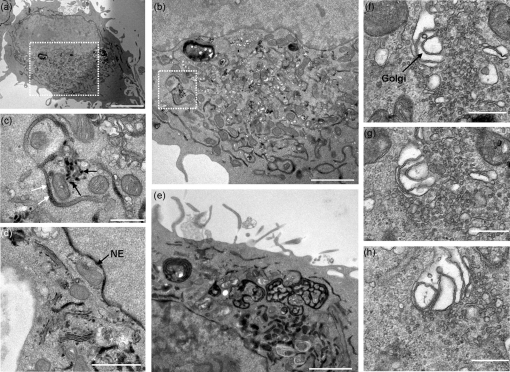
Fig. 8.
FCV-infected CRFK cells expressing the ER-retained HRPKDEL protein (black) exhibit characteristic signs of calicivirus infection and show correlation to transient protein studies in 293T cells. TEM of CRFK cells expressing the HRPKDEL protein and infected with FCV (Urbana) at an m.o.i. of 5. (a) Low magnification image of FCV-infected cells showing enlarged and circularized morphology, organelle polarization, vesicle formation and reduced HRPKDEL signal. (b) Enlarged image of the boxed area in (a). (c) Enlarged image of the boxed area in (b) showing ER dilation (white arrows), an associated loss of ER resident HRPKDEL signal and putative budding/release of HRPKDEL from the ER (black arrows). (d) High magnification image of FCV-infected CRFK cells showing accumulation of HRPKDEL in all stacks of the Golgi apparatus. (e) Infected CRFK cells exhibiting membranous ER structures analogous to those seen in 293T cells expressing FCV p30. (f–h) A proportion of infected CRFK cells exhibited dilation in stacks of the Golgi apparatus. Images are representative images taken from a Z stack series through the cell. NE, Nuclear envelope. Bars, 5 μm (a), 2 μm (b, e), 500 nm (c, f, g, h), 1 μm (d).

