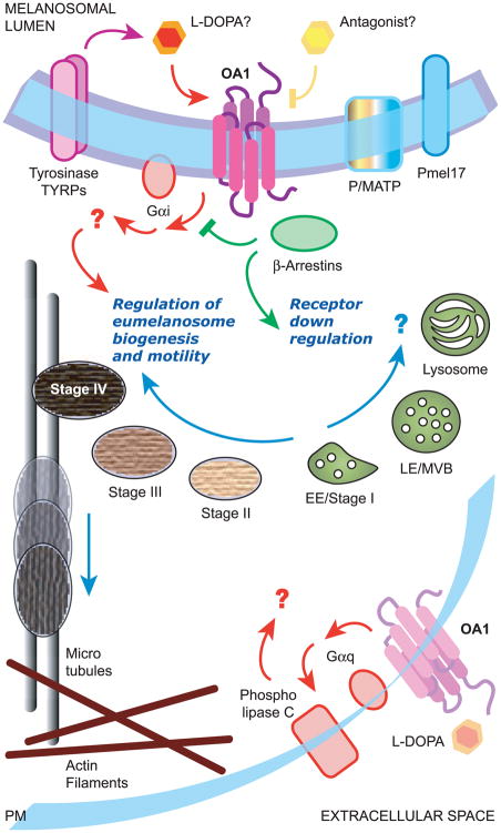
Figure 3.
Schematical overview of the signaling pathway mediated by OA1. Endogenous OA1 resides on the membrane of late endosomes/lysosomes and melanosomes, where it could function as a “sensor” of melanosome maturation and be activated by molecules belonging to the melanin biosynthetic pathway, possibly L-DOPA. It appears coupled to Gi proteins, through which it regulates proper biogenesis of melanosomes and restrains their transport toward the cell periphery by still unknown effectors. While a role in melanosome biogenesis and transport is supported by numerous lines of evidence, it is unclear whether the lysosomal localization of endogenous OA1 in melanocytes has a functional or downregulation role. In addition, a minor fraction of endogenous OA1 might travel to the cell surface of RPE cells, where it could become activated by L-DOPA and in turn trigger a Gq-mediated signaling pathway eventually leading to the secretion of neurotrophic factors. In contrast, exogenous OA1 overexpressed in non-melanocytic cells can be substantially missorted to the plasma membrane, downregulated by arrestins and coupled to Gq proteins, leading to phospholipase C activation and inositol phosphate or intracellular calcium increase (not shown). PM, plasma membrane; EE, early endosomes; LE, late endosomes; MVB, multivesicular bodies.

