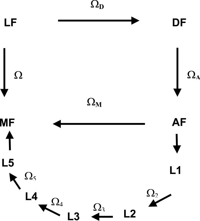
Figure 2.
Reference frames considered in this work.
LF: laboratory frame, with the ZL axis parallel to the magnetic field B0.
DF: PAS of the rotational diffusion tensor of the protein.
AF: frame of the amino acid residue, with the origin on the Cα bringing the nitroxide side-chain, the zA-axis perpendicular to the plane of the N-Cα-CO atoms and the xA-axis on the plane containing the zA-axis and Cα-Cβ bond.
Li: local frame, with the zLi axis parallel to the i-th chain bond and xLi in the plane of the preceding chain bond for the eclipsed configuration with χi−1 = 0° .
MF: magnetic frame, with the origin on the nitroxide N nucleus, the zM-axis along the N-pz orbital and the xM-axis parallel to the N-O bond.

